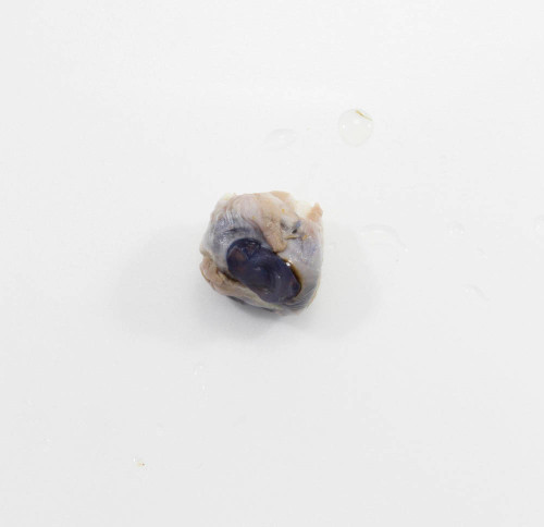
Sheep eye dissection offers a lower-cost alternative to cow eye dissection. Like a cow eye, a sheep eye is similar to the human eye, making it a popular dissection specimen.
Use a scalpel/dissecting razor blade and dissecting pan to explore the amazing ways in which a sheep's eyeball works. Examine the different parts of the eye, such as the cornea at the front of the eye, the front and back side of the iris (as well as the center of the iris), the oblong shape of the sheep pupil, the sclera, the vitreous humor and aqueous humor, the optic disc or blind spot (where the optic nerve connects at the back of the eye), extrinsic muscle bundles, the underlying choroid layer, the hemispheres of the eye, the ciliary body, other connecting muscles, veins, fatty tissue, and other sheep eye parts and eye structures.
Learning about the various eye functions and parts of a sheep's eye - by studying both the parts of the internal eye and external eye - will give students a memorable, hands-on life science experience. After seeing each part of the eye themselves, students can better understand how an eye works (e.g. how it focuses light).
Dissect this plain preserved specimen of an adult sheep eye with an individual student, or order several to conduct the dissection in a larger group!
Note: Specimens are initially preserved with a formaldehyde solution, the best animal tissue fixative. The formaldehyde is then displaced first with water and finally with a glycol solution to produce a moist, low-fume specimen which will not decay over time.
Important Notice: For orders of 10 or more, there is a chance these specimens could be shipped in a 10-pack. If you require specimens to be individually packaged, please contact our customer service at (406) 256-0990.
HST Specimen Guarantee
In sealed, original packaging, our preserved specimens are guaranteed to remain fully preserved and free of decay for 12 months from the date of purchase.
Once the original package is opened, use specimen within one month. For best results, observe the following storage procedures:
- Store specimen in heavy-duty, zip-lock bags to minimize drying between dissections.
- Specimen will slowly dry out or become contaminated in zip-lock bags; add a teaspoon of Specimen Holding Fluid to retain moisture.
- Freezing or refrigeration is not necessary and may damage fragile tissues.
 Warning

